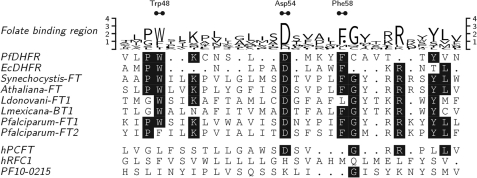
FIGURE 1.
Folate binding region. Residues 45–66 of P. falciparum DHFR aligned to E. coli DHFR (residues 19–36) as in (46) are shown. Other sequences are as in supplemental Figs. S1 and S3. Initial alignment was performed and prepared as in supplemental Fig. S3. Local alignment was optimized manually. Residues that interact with folate substrates and antifolates are marked at the top as Trp-48, Asp-54, and Phe-58 (46). Trp-48 is equivalent to the W100 in PfFT1, which is absent in PfFT2.

