FIGURE 9.
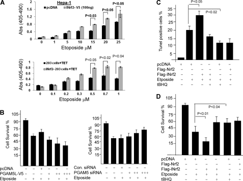
Nrf2-INrf2-PGAM5-Bcl-xL and cellular apoptosis. A, overexpression of INrf2 leads to an increase in etoposide-mediated apoptosis. Hepa-1 cells were transfected with pcDNA or INrf2-V5 plasmids and treated with etoposide for 48 h (top). Control 293 and INrf2-293 cells were treated with tetracycline and then treated with the indicated concentration of etoposide for 48 h (bottom). Transfected/treated Hepa-1 and treated 293/INrf2-293 cells were analyzed for apoptotic cell death by a DNA fragmentation assay. The cytoplasmic histone-associated DNA fragments (mono- and oligonucleosomes) were quantified using the Cell Death Detection ELISA kit (Roche Applied Science) and plotted (top and bottom). B, alterations in PGAM5 levels lead to an inverse relationship with cell survival. Hepa-1 cells were plated at a density of 5000 cells/well in 24-well plates and transfected with different concentrations of PGAM5L-V5 constructs (0.5, 1, and 2 μg) (left) or transfected with different concentration of siRNA against PGAM5 (25, 50, and 100 nm) for 24 h (right). The cells were then treated with DMSO or etoposide (20 μm) for 36 h. Cells were incubated with fresh MTT solution for 2 h at 37 °C, and absorbance at 570 nm was measured. The experiment was repeated three times. Each data point represents a mean ± S.D. and is normalized to the value of the corresponding control cells. C, TUNEL assay. t-BHQ treatment reduces etoposide-mediated apoptosis. Hepa-1 cells were transfected with FLAG-INrf2 or FLAG-Nrf2 and treated with etoposide for 48 h. One set of cells was further treated with t-BHQ for an additional 24 h, cells were fixed and permeabilized, and the TUNEL assay was performed. TUNEL-positive cells were observed under a fluorescence microscope, quantified, and plotted. The data are represented as the mean ± S.D. from two experiments. D, cell survival assay. Hepa-1 cells were plated at a density of 5000 cells/well in 24-well plates and transfected with FLAG-INrf2 or FLAG-Nrf2 and treated with DMSO or etoposide for 48 h and t-BHQ for 24 h. Cells were incubated with fresh MTT solution for 2 h at 37 °C, and absorbance at 570 nm was measured.

