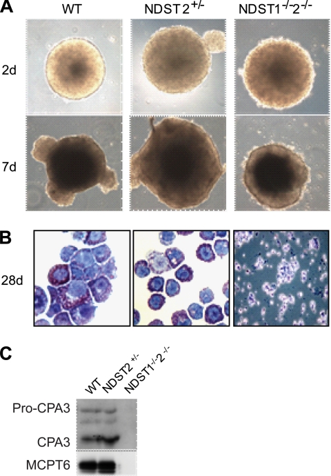
FIGURE 4.
ES cell-derived MCs. A, EBs of different genotypes cultured in suspension photographed at day 2 and at day 7 after plating. B, May Grünwald/Giemsa staining of WT and NDST2+/− MCs, and NDST1−/−2−/− cells from EB outgrowths placed on cytospin glasses. C, MC protease expression analyzed by Western blotting as described under “Experimental Procedures.”

