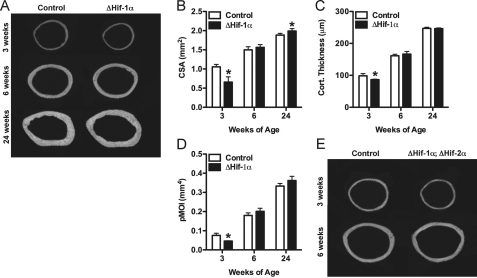
FIGURE 1.
Mice lacking Hif-1α in osteoblasts acquire increased cortical bone. A, representative microCT images illustrate cortical bone structure at the femoral mid-diaphysis in control and ΔHif-1α mice at 3, 6, and 24 weeks of age. B–D, bar graphs show quantification of the tissue cross-sectional area (CSA) (B), cortical thickness (Cort. Thickness) (C), and polar moment of inertia (pMOI) (D). E, representative microCT images from the femoral mid-diaphysis in control and ΔHif-1α;ΔHif-2α double mutants. Data are plotted mean ± S.E. with at least five mice being examined per genotype. *, p < 0.05

