Figure 2.

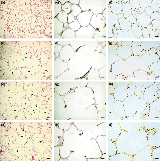
Photomicrographs of lung parenchyma histology 50 days after instillation of porcine pancreatic elastase (PPE, right lung) or saline (left lung). Haematoxylin–eosin staining (a–d) magnification ×100 (bar 200 μm): (a) saline-instilled lung showing the maintenance of the lobular and acinar architecture; (b) PPE-instilled lung from a control rat; note the hyperdistension of the alveolar ducts (*) with rupture of the alveolar septa (arrows); (c) PPE-instilled lung from a diabetic rat with distortion of the acinar architecture because of hyperdistension of the alveolar ducts (*) associated with the rupture of alveolar septa (arrows); and (d) PPE-instilled lung from insulin-treated diabetic rat showing similar histological pattern to PPE-instilled lung from control rat. Resorcin–fuchsin staining (e–h) magnification ×400 (bar 50 μm): (e) saline-instilled lung showing integrity of the elastic component in the alveolar walls, contrasting with disruption and thickening distribution of the fibres along the alveolar wall of PPE-instilled lung from a control rat (f); and decrease in PPE-instilled lung from a diabetic rat (g); note the restoration of the elastic system fibres pattern in PPE-instilled lung from insulin-treated diabetic rat (h). Picrosirius staining under polarized light (i–l) magnification ×400 (bar 50 μm): (i) note the weak orange-green birefringence of collagen fibres in saline-instilled lung compared to (j) PPE-instilled lung from a control rat; (k) PPE-instilled lung from a diabetic rat; and (l) PPE-instilled lung from insulin-treated rat.
