
Figure 6.
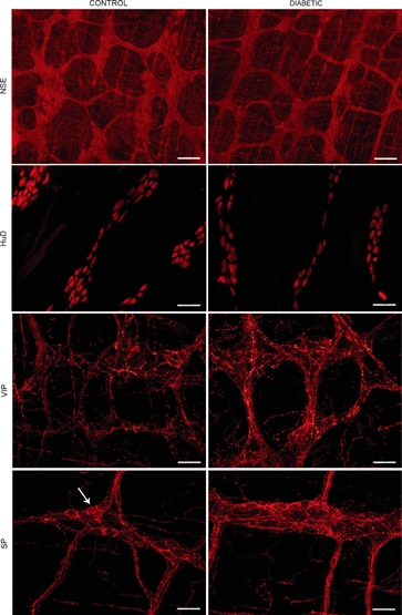
Immunohistochemistry on longitudinal muscle-myenteric plexus (LMMP) whole-mount preparations of ileum and colon of control and diabetic mice. Representative micrographs of neuron-specific enolase immunostaining, demonstrating the ganglionated myenteric network in the colon, which is less dense in diabetic mice; HuD immunostaining, showing immunoreactive neuronal somas of the ileal MP, with no staining of nerve fibres. Myenteric neurons were less numerous in samples from diabetic animals; VIP immunofluorescence in the colonic MP, demonstrating positivity primarily confined to neuronal processes and denser staining in samples from diabetic mice; SP immunofluorescence in the ileal MP, showing dense networks of fluorescent fibres, which are more abundant in diabetic animals. Note scattered faintly labelled neuronal somas (arrow).