
Figure 7.
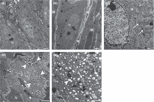
Representative transmission electron micrographs of the ileal and colonic myenteric plexus (MP) of control (a–c) and diabetic mice (d–e). a) Normal-appearing myenteric ganglion containing three neurons (N) and two enteric glial cells (G). (b) Neuromuscular arrangement in the MP and its close relationship with interstitial cells of Cajal (ICC) of the MP. A myenteric neuron (N) is surrounded by ICC processes (asterisks), which are in turn surrounded by smooth muscle cells (M). (c) Detailed view of the nuclear and perinuclear region of a normal-appearing myenteric neuron. Note rough endoplasmic reticulum (asterisk) and numerous mitochondria (arrows). (d) Abnormal myenteric ganglion. A neuron with preserved structure is shown (N), and at least two adjacent neurons exhibit severe vacuolization of the perikaryon (arrowheads). (e) Closer view of a myenteric neuron with signs of moderate-severe cell injury. Empty vacuoles corresponding to dilated rough endoplasmic reticulum (arrowhead), Golgi complex and swollen mitochondria with disrupted cristae (arrows) are seen.