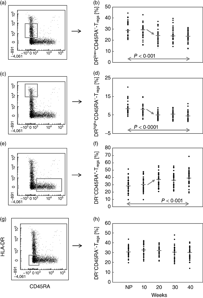
Fig. 3.

Detection of the percentages of the DRlow+CD45RA-, DRhigh+CD45RA-, DR-CD45RA- and DR-CD45RA+ Treg subsets within the total CD4+CD127low+/−CD25+forkhead box protein 3 (FoxP3+) Treg cell pool during the normal course of pregnancy. The figure shows the dot-blots of the gated DRlow+CD45RA-, DRhigh+CD45RA-, DR-CD45RA- and DR-CD45RA+ Treg subsets and their quantitative changes during the normal course of pregnancy. The diagram presents the individual and median data obtained for non-pregnant (NP) and healthy pregnant women. The percentage of the DRlow+CD45RA- Treg subset (a,b) and of the DRhigh+CD45RA- Treg subset (c,d) decreased significantly during the 10th and 20th weeks of gestation. In contrast, the percentage of the DR-CD45RA+ Treg subset increased during the same time (e,f). The percentage of the DR-CD45RA- Treg subset did not show any significant changes during the whole course of pregnancy (g,h).
