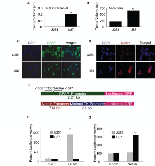
Figure 1: Tumorigenicity of GBM cells correlates with expression of Nestin and GFAP.
(A) Rat right frontal lobes were implanted with 5×10⁵ U251 or U87 cells in 5 µl PBS. After 3 weeks, tumor volumes were determined by pixel imaging analysis. (B) The right flanks of nude mice were injected (s.c.) with 2.5×10⁶ U251 or U87 cells in 100 µl PBS (mixed with 100 µl matrigel). After 5 weeks, tumor volumes were calculated using the formula: volume = width2 × length × 0.4. U251 or U87 cells grown on coverslips were fixed, permeabilized and stained for (C) GFAP (green) and (D) Nestin (red) using respective primary antibodies and secondary antibodies conjugated with Alexafluor488 and Alexafluor568 respectively. (E) Schematic of luciferase (Luc) reporter constructs: GFAP-Luc constitutes a 2.21 kb human GFAP promoter containing a GAS element (−1558 to −1547) in pGL3-Basic vector (upper panel) and Nestin-Luc constitutes a 714 bp enhancer fragment form the second intron of the human Nestin gene cloned upstream of an 81 bp minimal thymidine kinase (TK) promoter driving the luciferase gene in the vector, TP222 (lower panel). Activities of the GFAP promoter (F) and the Nestin enhancer (G) were determined at 72 h post transfection of U251 and U87 cells (1×10⁶) with indicated reporter plasmids (or empty vectors) by luciferase assay. For (A) & (B), each value represents mean ± SE of 5 individual animals of each group. Normalized percent luciferase values for (F & G) are plotted as mean ± SE (n = 3). ** indicates p < 0.01

