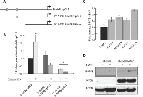
Figure 3: MYCN transcriptionally activates B-MYB MRNA and protein.
A) Schematic representation of three reporter vectors containing fragments of the B-MYB promoter: B-MYBp-pGL2 (from position −102 to −709, relative to the transcription start site); 5’-Δ360 (from position −102 to position −360); 5’-Δ240 (from position −102 to −240). B) Quantifications of luciferase assays showing the activity of the B-MYB promoter segments in the presence or absence of exogenously expressed MYCN. Error bars indicate standard deviations and the asterisk indicates statistically significant differences [* = p < 0.001] with respect to the levels of B-MYBp-pGL2 promoter activity, which was arbitrarily set to 1. C) Real time PCR analysis of B-MYB expression in 293 cells transiently transfected with a MYCN expression vector. The expression levels of four independent MYCN transfectants relative to cells transfected with empty vector are shown. Error bars indicate standard deviations obtained from triplicate assays. D) Western blot analysis showing enhanced expression of B-MYB caused by activation of MYCN in SKANAS::MYCN(ER) cells. (+ or −) 4-OHT indicates that the cells were cultured in the presence or absence of 4-hydroxytamoxifen, respectively.

