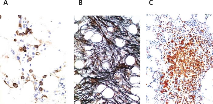
Figure 2. Diagnostic potential of histomorphological features in MDS and delineation of MDS subtypes.

A: Immunohistochemistry reveals an increase in CD34+ precursor cells in a patient with hypoplastic MDS. Note focal clustering of CD34+ precursor (blast) cells in the bone marrow section. B: The fibrotic form of MDS (MDS-F) as evidenced by Gömöri's silver stain (grade III). C: MDS associated with systemic mastcytosis (SM-MDS) as evidenced by staining the bone marrow section for mast cell tryptase. Note the compact infiltrate of spindle shaped tryptase-positive mast cells in this patient.
