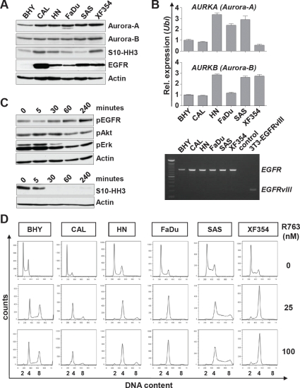
Figure 4. Expression and activity of Aurora kinases and EGFR in SCCHN cell lines.
(A) Six SCCHN cell lines were assessed by immunoblotting for the expression of Aurora-A and Aurora-B, for Aurora kinase activity measured by Histone H3 phosphorylation at serine10 (S10-HH3), and for EGFR protein levels. (B) Upper panel: AURORA-A and AURORA-B transcript levels were assessed by real-time qRT-PCR. Shown is the relative expression normalized to the expression of Ubiquitin. Lower panel: Expression of EGFR analyzed by RT-PCR. None of the SCCHN cell lines express the EGFRvIII mutant. Transiently transfected NIH-3T3 cells expressing EGFRvIII (3T3-EGFRvIII) were included as a control. (C) Upper panel: CAL cells were treated with 200 nM Cetuximab for the indicated time and assessed by immunoblotting for suppression of EGFR downstream target phosphorylation. Lower panel: Treatment of FADU cells with 5 nM Pan-Aurora kinase inhibitor R763 for the indicated time. The activity of Aurora kinases was assessed by immunoblotting for S10-HH3. (D) SCCHN cell lines were treated for 24 hr with R763 at the indicated concentrations or carrier alone (0 nM). The representative histograms show the DNA content assessed by propidium iodide (PI) staining.

