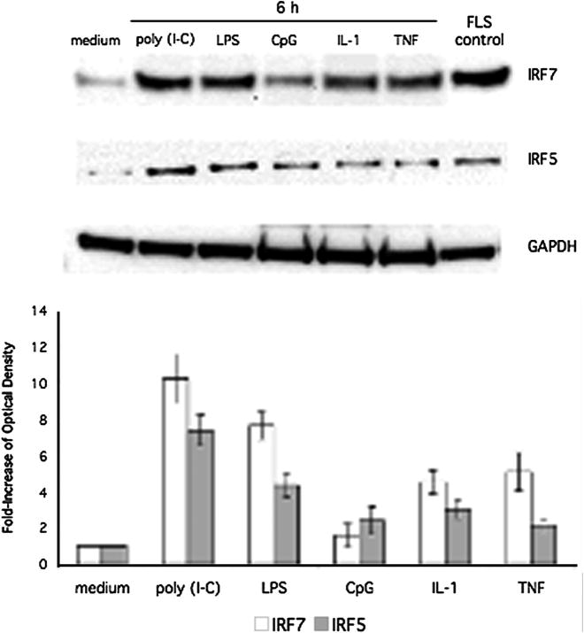
Figure 1.
Western blot analysis of IRF5/7 induction. THP-1 cells were stimulated for 6 h with poly (I-C), LPS, CpG, IL-1, or TNF. Lysates were then analyzed by Western blot using anti-IRF5, anti-IRF7, and anti-GAPDH antibodies. Lysate from poly (I-C) stimulated fibroblast like synoviocytes (FLS) was used as a positive control. Stimulation with poly (I-C), LPS, IL-1, and TNF showed significant induction of IRF7 and IRF5. Poly (I-C) showed the most significant increase (10.26-fold ± 1.36 and 7.39-fold ± 0.85; n = 3 respectively). Top panel shows a representative Western blot, and the bottom panel shows combined quantification of protein expression by densitometry for three independent experiments.

