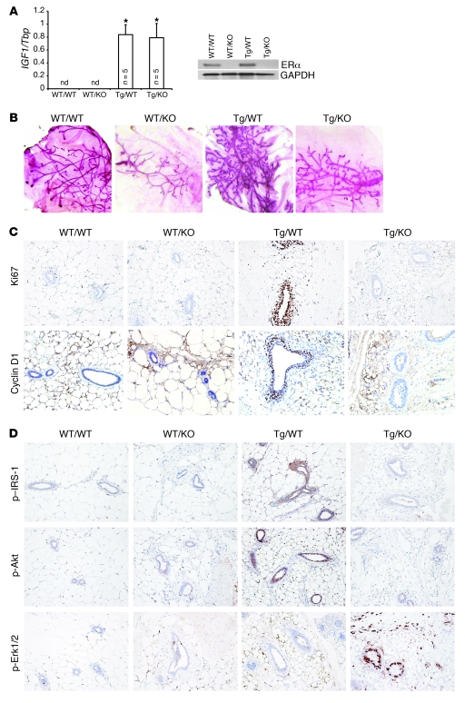
Figure 6. Analysis of mammary glands from 4- to 5-week-old bitransgenic BK5.IGF-1 × ERKO mice, which overexpress IGF-1 but lack ERα.
Crossing the two models generated mice of 4 genotypes, designated as follows: IGF-1 WT/ERα WT (WT/WT), IGF-1 WT/ERKO (WT/KO), BK5.IGF-1 Tg/ERα WT (Tg/WT), and BK5.IGF-1 Tg/ERKO (Tg/KO) mice. (A) Evaluation of transgene expression by qPCR (left panel) showing that human IGF1 is expressed exclusively in BK5.IGF-1 Tg mice. Western blot analysis of ERα expression shows that the receptor is not expressed in glands from ERKO mice (right panel). The bar graphs represent the mean ± SD of separate experiments, as indicated by n in the figure. Statistical analysis performed using Student’s t test; *P < 0.05 versus WT/WT glands. (B) Representative whole mounts of mammary glands (original magnification, ×1), showing that Tg IGF-1 does not rescue the ERKO phenotype. (C) Ki67 and cyclin D1 immunostaining in mammary glands from mice with each of the 4 bitransgenic genotypes (original magnification, ×20 and ×40, respectively), showing increased proliferation and cyclin D1 expression in the mammary epithelium of Tg/WT, but not Tg/KO, mice. (D) Immunohistochemical localization of p–IRS-1, p-Akt, and p-Erk1/2 in paraffin-embedded sections of mammary glands from mice with the 4 genotypes (original magnification, ×20). nd, not detectable.

