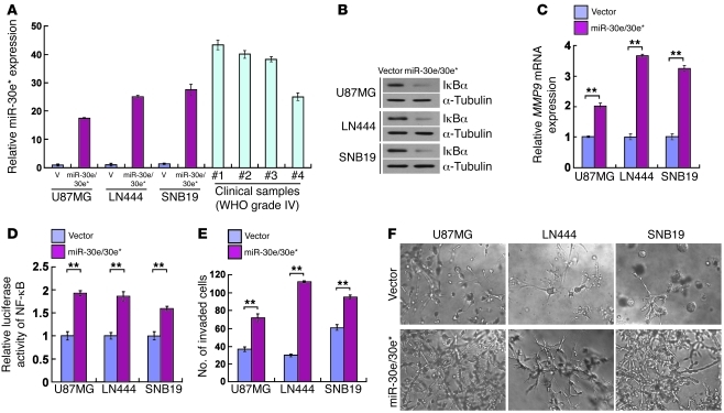
Figure 6. miR-30e/30e* induces invasiveness of glioma in vitro.
(A) Real-time PCR analysis of miR-30e* expression in the indicated cells and 4 WHO grade IV glioma samples compared with that in U87MG cells. Transcript levels were normalized by U6 expression. (B) Western blot analysis of IκBα protein in glioma cells transduced with a retroviral vector to express miR-30e/30e*. α-Tubulin was detected as the loading control. (C) Expression of MMP9 mRNA was upregulated in miR-30e/30e*–transduced cells. Expression levels were normalized by GAPDH. (D) The relative NF-κB reporter activity increased in miR-30e/30e*–transduced cells. (E) Quantification of indicated invaded cells in the TMPA with Matrigel. (F) Representative micrographs of indicated cells cultured in the 3D spheroid invasion assay. Original magnification, ×200. Experiments in A–F were repeated at least 3 times, with similar results. **P < 0.01.

