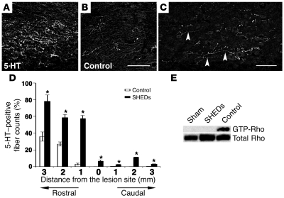
Figure 4. SHEDs regenerate 5-HT fibers and inhibit SCI-induced activation of Rho GTPase.
(A–D) Representative images (A–C) and quantification (D) of serotonergic raphe axons stained with 5-HT mAb in the sagittal sections of the transected SC. A large number of 5-HT axons penetrated the scar tissue of the rostral stump in the SHED-transplanted SC (A), while only a few did in the control transected SC (B). (C) 5-HT–positive boutons were in contact with neurons of the caudal stump. Arrowhead indicates 5-HT–positive fiber extended beyond the epicenter. Quantification of regenerated 5-HT axons (D) was carried out as described in Figure 2D, except the y axis indicates the percentage of 5-HT axons compared with that in the sham-operated SC. Data represent the mean ± SEM. *P < 0.01 compared with SCI models injected with PBS. Scale bars: 50 μm (B and C). (E) SCI-induced Rho GTPase activation 7 days after SCI was suppressed by engrafted SHEDs. The level of active Rho in lysate from the samples indicated at the top (sham-operated, control, and SHED-transplanted) was examined by RBD pull-down assay.

