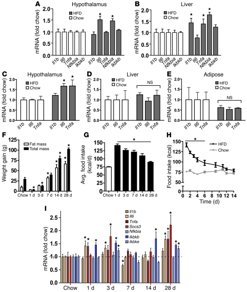
Figure 1. Time course of hypothalamic inflammation after the onset of HFD feeding.
(A and B) Quantification of mRNA encoding proinflammatory cytokines (Il1b, Il6, Tnfa) and NF-κB pathway genes (Nfkbia, Ikbkb) in (A) hypothalamus and (B) liver of rats fed either standard chow (white bars) or HFD (gray bars) for 20 weeks (n = 6/group). *P < 0.05 versus chow-fed controls. (C–E) Effect of 4 weeks of HFD feeding (gray bars) on proinflammatory cytokine gene expression in rat (C) hypothalamus, (D) liver, and (E) white adipose tissue compared with that in chow-fed controls (white bars) (n = 6/group). *P < 0.05 versus chow-fed controls. (F) Total weight gain (black bars), fat mass gain (white bars), and (G) average (avg) daily food intake (kcal/d) of rats (n = 6/group) fed chow for 2 weeks or HFD for up to 28 days. *P < 0.05 versus chow-fed controls. (H) Comparison of daily food intake (kcal) in rats (n = 6/group) fed chow (gray) or HFD (black) for 14 days. *P < 0.05 versus chow-fed controls. (I) Time course of induction of mRNA encoding inflammatory mediators, including proinflammatory cytokine (Il1b, Il6, Tnfa), cytokine pathway (Socs3), and NF-κB pathway (Nfkbia, Ikbkb, Ikbke) gene expression in the hypothalamus of rats fed chow or HFD for up to 28 days (n = 6/group). All mRNA species were quantified relative to 18S and Gapdh housekeeping gene expression (by ΔΔCT method) and presented as fold change relative to chow-fed controls [fold chow]. The dashed line in I represents the level of expression equal to chow-fed controls. *P < 0.05 versus chow.

