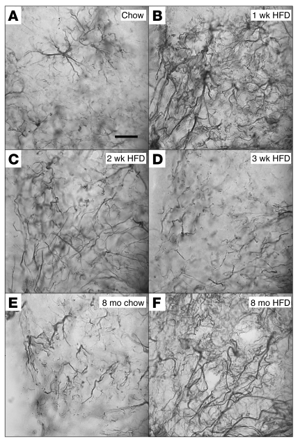
Figure 5. Effect of HFD feeding on ARC astrocyte morphology.
High-magnification (original magnification, ×100) examination of astrocyte processes by GFAP immunohistochemistry of sections through mouse ARC. (A) Astrocyte processes in the ARC of mice fed chow remain separated into discrete areas. (B) One week of HFD feeding is accompanied by the apparent formation of a syncytium of astrocytic processes. (C) This astrocyte response is partially resolved by 2 weeks of HFD feeding, with only a few scattered overlapping processes, and, (D) by 3 weeks of HFD, glial morphology appears to be fully normalized. (E) Mice fed chow for 8 weeks show increased astrocyte number but no overlap of processes. (F) Mice fed HFD for 8 months exhibit severe astrocytosis suggestive of syncytium formation. Scale bar: 10 μm.

