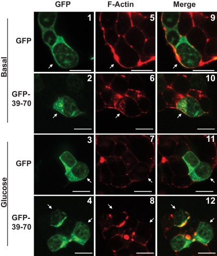
Fig. 4.
GFP-39–70 does not disrupt normal glucose-induced actin remodeling. MIN6 cells were plated on glass coverslips and transfected to express GFP or GFP-39–70 proteins (green) were preincubated in MKRBB for 2 h and left unstimulated (top panels 1 and 2, 5 and 6, and 9 and 10) or stimulated with 20 mm glucose for 5 min (bottom panels 3 and 4, 7 and 8, and 11 and 12). Cells were fixed, permeabilized, and cortical F-actin staining detected by rhodamine-phalloidin staining (red). Cells in clusters containing both nontransfected and GFP-transfected cells were imaged at the midplane of the cluster by single channel scanning confocal microscopy to determine subcellular distribution of the GFP proteins (panels 1–4) and F-actin remodeling (panels 5–8). Merged GFP and F-actin images are shown in panels 9–12 to permit comparisons between nontransfected and transfected cells. Scale bar, 10 μm. Data shown are representative of three independent sets of experiments.

