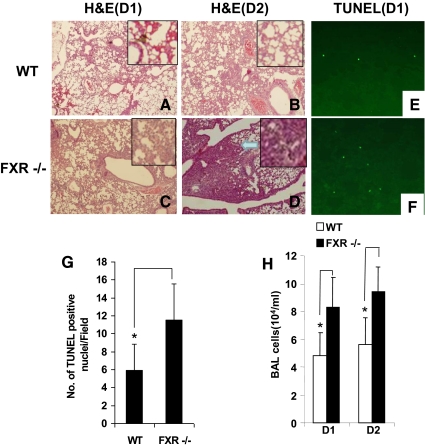
Fig. 2.
FXR−/− mice are more susceptible to LPS-induced ALI. A–D, Histologic analysis revealed elevated neutrophilic alveolar and interstitial infiltration by d 1 and 2 after LPS inhalation in FXR−/− mice, with increased interstitial thickening and mixed cellular infiltration on d 2 in the lungs of FXR−/− mice compared with the control WT. Arrow indicates increased interstitial thickening and mixed cellular infiltration. E and F, Representative micrographs of terminal deoxynucleotidyl transferase 2-deoxyuridine, 5-triphosphate nick end labeling (TUNEL) staining of WT and FXR−/− lung tissue after LPS challenge. Lung sections of WT (E) and FXR−/− (F) mice collected 24 h after LPS challenge were stained with FITC-conjugated TUNEL to identify apoptotic cells. G, Number of the TUNEL-positive cells on D1 after LPS treatment. H, Total BAL cell counts of WT and FXR−/− mice after treatment with LPS (cellular infiltration in the airways after exposure to LPS). *, P < 0.05. TUNEL staining revealed there were approximately twice as many positive cells in lung tissue from FXR−/− mice (n = 4) compared with WT control mice (n = 4). The number of cells is the average of at least three different fields from each mouse (E–G). In FXR−/− animals, LPS treatment also triggered an altered profile of leukocyte influx into the airspace, with a marked increased of BAL cells at d 1 (D1) (n = 6) and 2 (D2) (n = 6), as compared with the WT controls (H). H&E, Hematoxylin and eosin.

