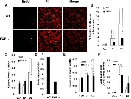
Fig. 4.
Defective lung regeneration in FXR−/− mice after LPS-induced ALI. A, Representative micrographs of BrdU immunostaining of WT and FXR−/− lungs after LPS-induced lung microvascular injury. Sections (5 μm) of lungs collected 48 h after LPS challenge were stained with FITC-conjugated anti-BrdU antibody to identify proliferating cells; nuclei were counterstained with propidium iodide. B, Quantification of BrdU-positive nuclei of the lung sections described in A at indicated time points after LPS challenge [d 1 (D1), n = 4; and d 2 (D2), n =4]. C, QRT-PCR analysis of Foxm1b mRNA levels in lungs collected from WT or FXR−/− mice at the indicated time points after LPS exposure. D, Fold change in Foxm1b gene transcription induction (D2/D0). E, Quantitative analysis of Cyclin D1 mRNA levels after treatment of WT or FXR−/− mice with LPS. F, Pulmonary permeability of WT or FXR−/− mice as a function of Evans blue dye extravasation at the indicated time points after treatment with LPS [d 1 (D1), n = 4 and d 2 (D2), n = 4]. *, P < 0.05. Con, Control; PI, propidium iodide.

