Figure 2.
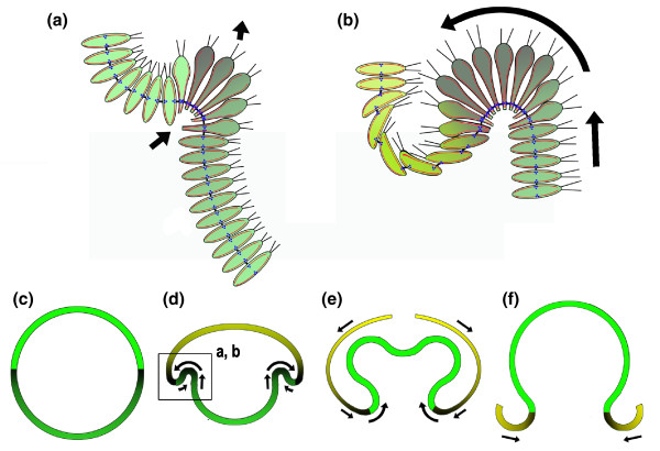
A sectional diagram illustrating aspects of the type B inversion in Volvox. (a,b) The initial steps in forming the subequatorial zone of cell wedging that progresses posteriorly and rolls or involutes (curved arrow) the posterior half into the anterior half are shown. (c-f) The location of this event in the context of the entire inversion is shown (box in (d)). The direction of the progression of zones of cell shape change is indicated by shading from dark to light (see text for discussion). Red, microtubules; blue, kinesins; black, cytoplasmic bridges. Based on [7-9].

