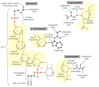
Figure 1. Schematic representation of glycosylation forms.
For each glycosylation type, the amino acid acceptor site is illustrated in balls and sticks: N-glycosylation (asparagine residue), O-glycosylation (serine residue), C-mannosylation (tryptophan residue), and glycosylphosphatidylinositol (GPI) anchor (C-terminal protein residue). Small balls colored in grey, red, blue, and orange represent carbon, oxygen, nitrogen, and phosphorus atoms, respectively. Hydrogen atoms were not shown. The atoms involved in glycan linkage are indicated with rows. Glycan molecules are shown as sticks and highlighted with a yellow background color. The GPI molecule was divided into three parts: phosphoethanolamine, glycan core, and phosphatidylinositol. The glycan core is composed of one non-acetylated glucosamine (GlcN) and three mannose moieties. The long fatty acids contained in the phosphatidylinositol portion are indicated using waves.

