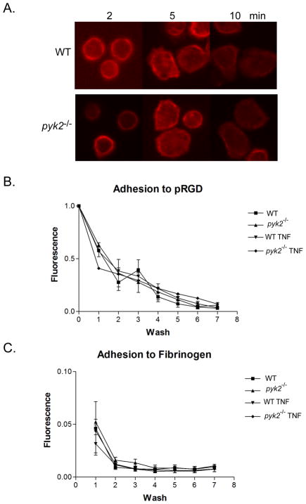
FIGURE 2.
Analysis of integrin-mediated adhesion in pyk2−/− PMNs. A, PMNs from WT or pyk2−/− mice were allowed to adhere to pRGD-coated coverslips and fixed at the indicated time points. The fixed cells were stained for actin (red). Images shown are representative of three independent experiments. B, Fluorescently-labelled bone marrow PMNs from WT or pyk2−/−mice were plated on pRGD coated wells and then exposed to a series of washes in a static adhesion assay. The decrease in fluorescence corresponding to the decrease in cell number was measured by fold-change from the baseline value was plotted over the series of washes. C, Following the assay design in B, PMNs from WT or pyk2−/− mice were allowed to adhere to fibrinogen coated wells for 15 minutes. Error bars represent ± SEM of triplicate samples. Data shown are pooled from 3 independent experiments.

