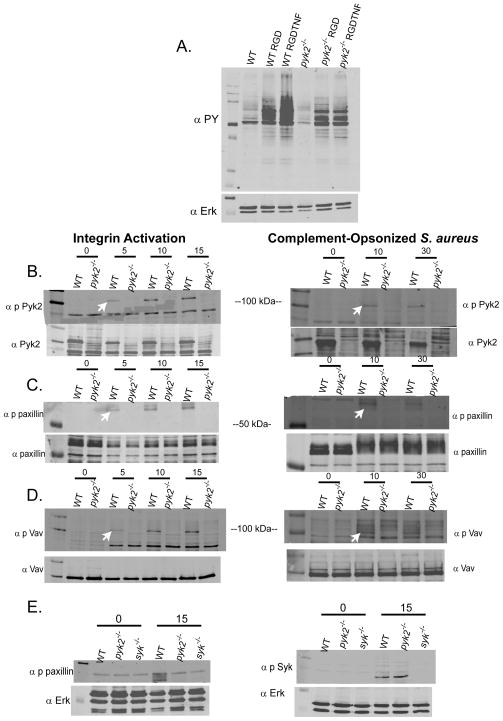
FIGURE 7.
Signal activation following integrin ligation in pyk2−/− PMNs. A, WT or pyk2−/−bone marrow-derived PMNs were plated on pRGD in the presence or absence of 100 ng/mL TNF for 15 minutes. PMNs in suspension were used as control. Lysates were immunoblotted for phosphotyrosine (4G10 antibody) with Erk1 used as an equal loading control. B–D, WT or pyk2−/− PMNs were activated on pRGD for 0, 5, 10 or 15 minutes (left panels) or by exposure to complement-opsonized S. aureus for 0, 10 or 30 minutes (right panels) and then lysed. Lysates were immunoblotted with Abs specific for phospho-Pyk2, phospho-paxillin or phospho-Vav. Arrows indicate the appropriate bands in each gel. Lower panels show the same lysates probed for total Pyk2, paxillin, and Vav as loading controls. Arrows indicate specific bands. Data are representative of 2–6 different experiments for each panel. E, Lysates from WT, pyk2−/− and syk−/− PMNs activated on pRGD for 15 minutes were probed for phospho-paxillin and phospho-Syk as indicated with Erk1 serving as an equal loading control.

