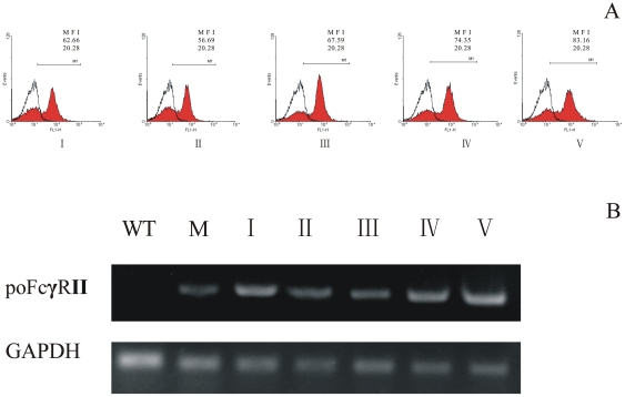
Figure 1. Binding of porcine IgG to poFcγRII on Marc-poFcγRII cells was monitored by flow cytometry.
(A). Heat-aggregated IgG was applied to Marc-poFcγRII cells (I–V: passage 1, 5, 10, 15 and 20), followed by washing and incubation with the FITC-conjugated goat anti-porcine IgG. Open graph shows untransfected wild-type Marc-145 cells; Expression of poFcγRII on Marc-poFcγRII cells was determined by RT-PCR (B). Total mRNA was prepared and cDNA was synthesized. This cDNA was then used as a template in polymerase chain reaction (PCR) with poFcγRII or porcine glyceraldehydes 3-phosphate dehydrogenase (GAPDH) specific primers. (lane I–V: passage 1, 5, 10, 15 and 20, lane WT: parent wild-type Marc-145, and lane M: Macrophages).

