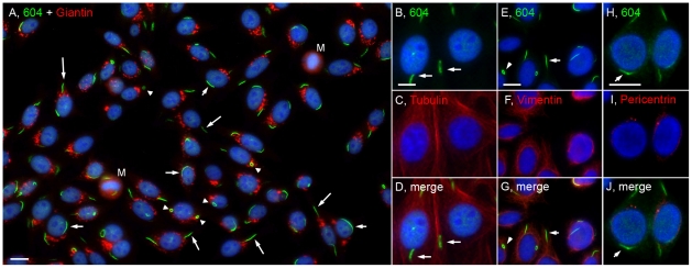
Figure 1. The distribution of cytoplasmic rods and rings was independent of the Golgi complex and centrosomes, and these structures were not enriched in tubulin or vimentin.
(A) Merged image of HEp-2 co-stained with human anti-RR prototype serum 604/Alexa 488 goat anti-human Ig (green) and rabbit anti-giantin (Golgi marker)/Alexa 568 goat anti-rabbit Ig (red). Rods are often presented adjacent (short arrows) or perpendicular (long arrows) to the nucleus while rings (arrowheads) are found either 1 or 2 to a cell. M, mitotic cell. HEp-2 cells were also co-stained with serum 604/Alexa 488 goat anti-human Ig (green, B,E,H) and different cytoplasmic markers using mouse anti-tubulin (C), anti-vimentin (F), anti-pericentrin (I), followed by Alexa 568 goat anti-mouse Ig (red). Nuclei were counterstained with DAPI (blue). Bar, 10 µm.

