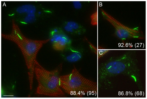
Figure 7. Inhibition of CTPS1-induced formation of RR in mouse primary cardiomyocytes, fibroblasts, and endothelial cells.
Mouse primary cardiomyocytes prepared together with fibroblasts and endothelial cells, were treated with 2 mM DON and cultured for 24 h. Cells were co-stained with human anti-RR serum IT2006 (green) and mouse anti-actinin monoclonal antibody (red). Nuclei counterstained with DAPI (blue). Actinin-positive cardiomyocytes (A, B, red) as well as actinin-negative fibroblast or endothelial cells (A, C) all show distinct rods. The percentage of cells with RR displayed is shown in the lower right corner with the total number of cells counted indicated in parentheses (A, all cells; B, actinin-positive cardiomyocytes only; C; actinin-negative fibroblast and endothelial cells). Bar, 10 µm.

