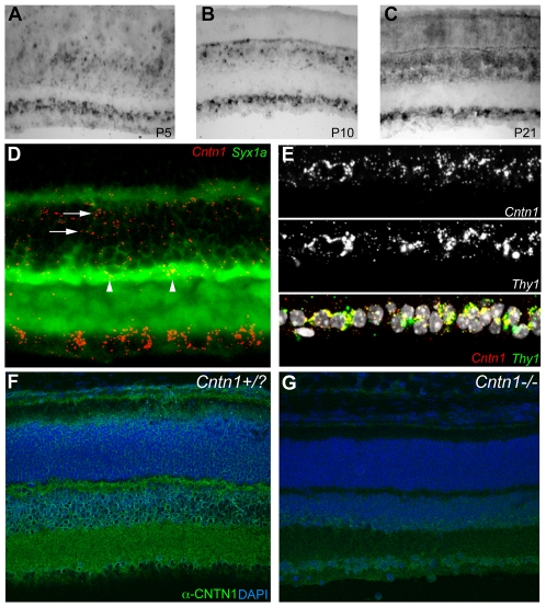
Figure 4. Expression and localization of Cntn1 in the retina.
A–C In situ hybridization for Cntn1 expression in the wild type mouse retina at P5 (A), P10 (B), and P21 (C). A subset of cells in the retinal ganglion cell layer (bottom) and inner nuclear layer are positive for Cntn1 expression. Photoreceptors in the outer nuclear layer (top) do not have signals above background. D) Double label in situ hybridization with Cntn1 and syntaxin1a at P21 demonstrates that some amacrine cells in the inner nuclear layer are positive for Cntn1 expression (arrowheads). Other Cntn1-positive cells are likely to be bipolar cells based on their position (arrows). E) In the retinal ganglion cell layer, a majority of cells expressing Cntn1 at P21 also express Thy1, a marker of ganglion cells. F) Immunolabeling of retinas with anti-CNTN1 antibodies at P14 revealed strong labeling of the synaptic plexiform layers, as well as immunoreactivity in the cellular layers, particularly the inner nuclear layer. G) Immunolabeling retinas from Cntn1 mutant mice revealed a marked reduction, but not an elimination of signal intensity in images collected with equivalent parameters.

