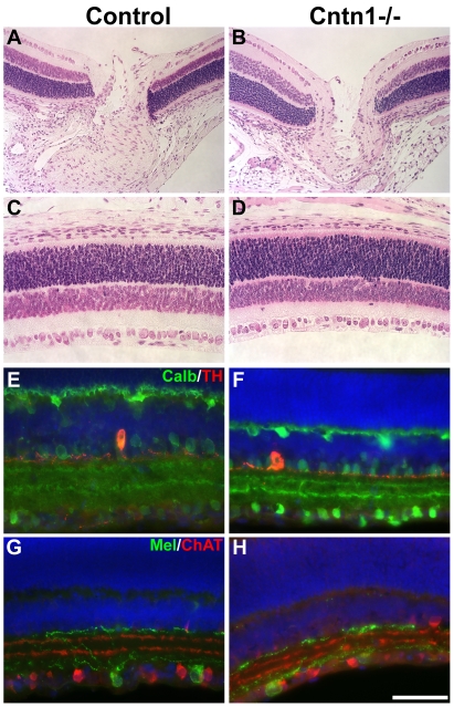
Figure 5. Normal retina development in Cntn1 mutant mice.
A,B) The retina and optic nerve head of wild type and Cntn1 mutant mice stained with H&E did not reveal any defects. C,D) More peripheral areas of the retina were also normal in the mutant. E,F) Staining for dopaminergic amacrine cells (red, anti-tyrosine hydroxylase) and horizontal cells and calbindin-positive amacrine cells (anti-calbindin, green), showed that cell bodies and dendritic arbors of these cells were in the appropriate anatomical location. G,H) Staining for intrinsically photoresponsive ganglion cells (anti-melanopsin, green) and starburst amacrine cells (anti-choline acetyltransferase, red) showed that these cells were also unaffected by the loss of Cntn1. The scale bar in H represents 145 µm in A–D and 72 µm in E–H.

