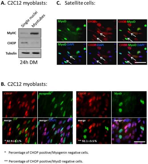
Figure 4. The expression of CHOP and MRFs is mutual exclusive.
(A) C2C12 cells were grown in DM for 24 hours and mononucleated cells were separated from myotubes by selective trypsinization. The two cell populations were subjected to Western blot for analyzing the expression of CHOP. (B) C2C12 myoblasts were grown in DM for 24 hours, and double stained with antibodies directed against CHOP and myogenin (left panel) or with antibodies directed against CHOP and MyoD (right panel). DAPI in blue, MyoD and myogenin in green and CHOP in red. Percentage of CHOP positive, myogenin negative and CHOP positive, MyoD negative relative to the total number of CHOP positive cells was calculated in three independent experiments. Mean values and standard errors are presented. Bar, 50 µm. (C) The expression of CHOP in primary satellite cells. To induce their differentiation, satellite cells were grown for 24 hours in GM medium. Cells were analyzed by immunostaining with anti-MyoD (green) and anti-CHOP (red) antibodies. DAPI staining is in blue. Arrows point at nuclei positive for CHOP staining and negative for MyoD staining. Bar, 50 µm.

