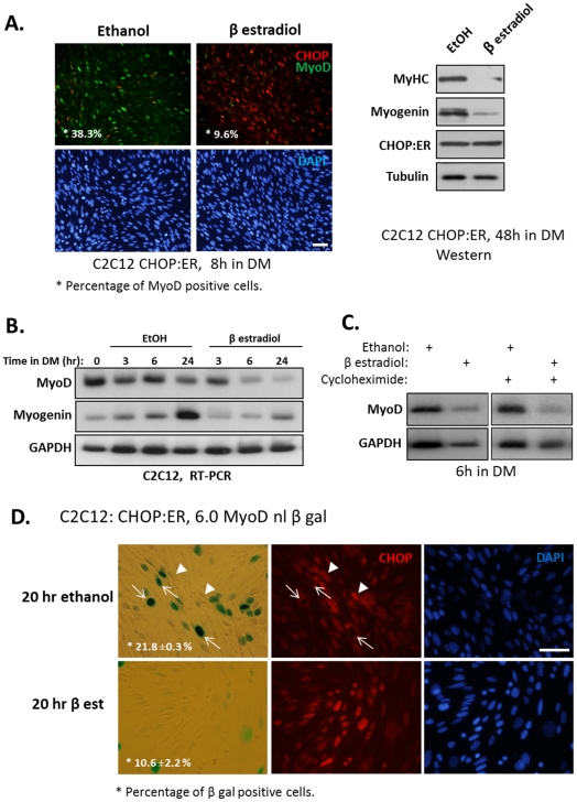
Figure 6. CHOP represses MyoD transcription.
(A) A C2C12 derived cell line expressing a chimera CHOP:ER protein was constructed as is described under “Materials and Methods”. (A) Myoblasts were grown in the presence of ethanol or β estradiol (0.1 µM) for 8 hours. Cells were immunostained using anti-MyoD and anti-CHOP antibodies. DAPI in blue, MyoD in green and CHOP in red. Percentage of MyoD-positive nuclei relative to the total number of nuclei was calculated. Bar, 50 µm. (left panel). In another experiment, cells were grown in the presence of ethanol or β estradiol (0.1 µM) for 48 hours and proteins were analyzed by Western blot (right panel). (B) The same cells as above were grown in DM in the presence of ethanol or β estradiol (0.1 µM) for the indicated time periods and mRNA levels of myod and myogenin were determined by semi-quantitative RT-PCR analysis. (C) The same cells as above were grown in DM and in the presence of ethanol or β estradiol for 6 hours in the absence or presence of cycloheximide added to cells one hour before the addition of ethanol or β estradiol. (D) A clone of the above cells (i.e., expressing CHOP:ER) with integrated MyoD reporter gene (6.0 MyoD -nl β Gal) was isolated. These cells were grown in the presence of ethanol or β estradiol for 20 hours. Nuclear expression of β Gal was identified by an enzymatic colorimetric assay, and the expression of CHOP by immunostaining. Arrows point at β Gal-positive nuclei that are CHOP negative. Percentage of β Gal-positive nuclei out of the total number of nuclei was calculated in two independent experiments. Mean values and standard errors are presented. Bar, 50 µm.

