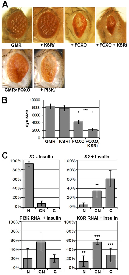
Figure 1. KSR is involved in the regulation of FOXO activity.
(A) Photomicrographs of adult eyes. Upper panel: UAS-KSRRNAi or UAS-FOXO or both were expressed in the developing eye with the GMR-GAL4 driver. Lower panels: GMR-GAL4+UAS-FOXO also expressing UAS-RNAi to deplete PI3K. (B) Quantification of the total area of affected eyes of the indicated genotypes measured in pixels from digital images using ImageJ. Error bars indicate standard deviation from measurement of at least 6 eyes for each genotype. Student's t-test: (***) p<0.001. (C) Quantification of the subcellular localization of transfected FOXO-GFP. S2 cells with GFP signal were classified into 3 groups according to FOXO localization (N: predominantly nuclear; CN: equal levels in cytoplasm and nucleus; C: predominantly cytoplasmic). Upper panels: compare unstimulated cells with cells stimulated with insulin (10 µg/ml, 30 min). Lower panels: Cells transfected with dsRNA to deplete PI3K or KSR and after 4 days, stimulated with insulin. Error bars represent standard deviation from 3 independent experiments. Fisher's exact test was used to assess the difference between insulin-stimulated S2 cells with and without KSR depletion: (**) p<0.01; (***) p<0.001.

