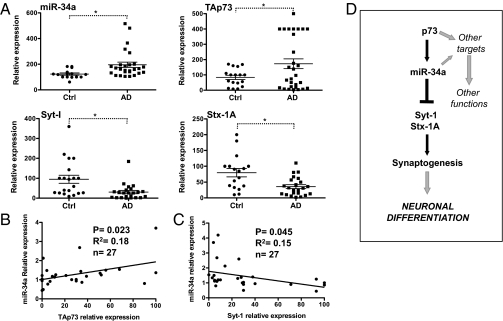
Fig. 4.
Expression of miR-34a, TAp73, and synaptic proteins in AD brain. (A) Total RNA was extracted from postmortem control and AD hippocampus. Expression of the indicated genes was evaluated by real-time PCR. Data represent mean ± SEM. *P < 0.05. (B) Pearson's test on individual AD samples show significant positive correlation between TAp73 and miR-34a expression. (C) A significant negative correlation between miR-34a and Syt-1. (D) Schematic model for the role of TAp73/miR-34a axis in developmental neuronal differentiation.

