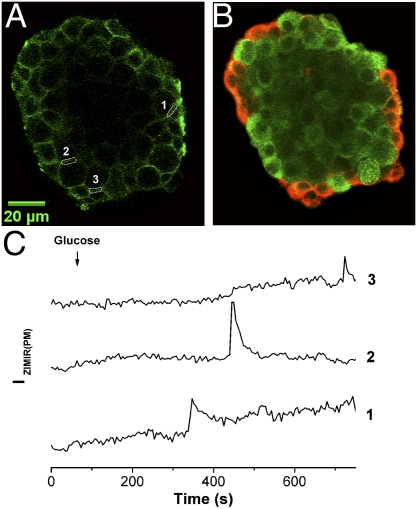
Fig. 7.
Islet β cells secreted insulin at both homologous and heterologous cell-cell contacts. (A) Confocal ZIMIR image of an islet before glucose stimulation. Three regions of interest (ROIs) along intercellular contacts are shown. (B) Confocal immunohistochemical image (red, glucagon; green, insulin) of the same focal plane of the same islet as in A. ROI-1 and ROI-3 correspond to α-β contacts, and ROI-2 corresponds to a β-β contact. (C) Time courses of ZIMIR fluorescence of ROI-1 through ROI-3. Scale bar, 20 μm.

