Fig. 2.
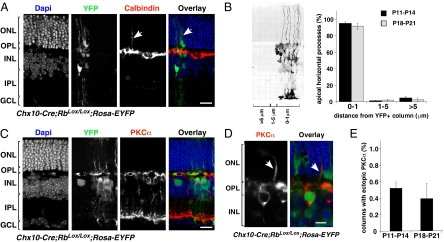
HC processes do not reorganize into their laminar position in Chx10-Cre;RbLox/Lox;Rosa-YFP mouse retinas. (A) Coimmunolocalization of YFP (green) and calbindin (red) in P21 Chx10-Cre;RbLox/Lox;Rosa-YFP mouse retinas. Apical processes (arrows) of calbindin-immunopositive HCs are associated with regions of Rb inactivation (green). (B) Association of apical processes was scored with respect to distance from a column of Rb-deficient cells, as indicated by YFP expression. Once the apical horizontal cell process was identified, the distance from the nearest YFP+ column of cells was measured, and processes were divided into a 0–1μm group (within the column of cells or touching the column on the edge); a 1–5 μm group (within approximately one cell body of the column); and a >5 μm group (beyond approximately one cell body of column). The histogram gives mean (± SD) of scoring from the six independent retinas at each stage. (C) PKCα-immunopositive rod bipolar cells did not extend dendrites into the ONL in Rb-inactivated (green) or wild-type regions at P21. (D) A rare ectopic PKCα dendrite in Chx10-Cre;RbLox/Lox;Rosa-YFP retinas (arrow) adjacent to a patch of YFP-expressing cells (green). (E) Histogram showing mean (± SD) scoring from the six independent retinas at each stage. Bip, bipolar cell; GCL, ganglion cell layer; HC, horizontal cell; INL, inner nuclear layer; IPL, inner plexiform layer; ONL, outer nuclear layer; OPL, outer plexiform layer. (Scale bars: 10 μm.)

