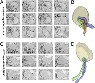
Fig. 3.
Serial sectioning and 3D reconstruction of ectopic and normal synaptic triads. (A) Electron micrographs of serial sections from the outer plexiform layer showing labeled bipolar and horizontal elements in a synaptic triad. (B) 3D reconstruction of micrographs in A with individual horizontal neurites shown in green and blue and the bipolar dendrite shown in orange. (C) Electron micrographs of serial sections from the ONL showing labeled horizontal elements in an ectopic synaptic triad. (D) 3D reconstruction of the micrographs in C with individual horizontal neurites shown in green and blue. No bipolar dendrites were observed.

