Fig. 2.
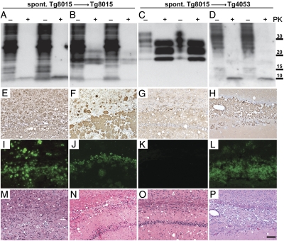
Characterization of spontaneously generated PrP(ΔGPI) prions upon passage in Tg8015/Prnp0/0 and Tg4053 mice. Different phenotypes were observed biochemically following parallel transmission of spontaneously generated prions into both Tg8015/Prnp0/0 and Tg4053 mice. (A–D; two examples shown in each case) Brain homogenates from spontaneously ill Tg8015/Prnp0/0 mice transmitted completely to Tg8015/Prnp0/0 mice within ∼200–300 d. We observed two different phenotypes: brains of two mice contained only the ∼10-kDa, PK-resistant band (A), whereas transmissions from two other brains resulted in a mixture of ∼10-kDa and 17- to 19-kDa PK-resistant fragments (B). In parallel experiments, we inoculated the same brain homogenates from Tg8015/Prnp0/0 mice into Tg4053 mice (C and D). Those brains that contained PK-resistant PrP of 10 kDa and 17–19 kDa upon inoculation to Tg8015/Prnp0/0 (B) transmitted efficiently to Tg4053 mice and resulted in a typical three-band PrPSc pattern (C). Conversely, a brain homogenate that resulted in only the ∼10-kDa fragment in Tg8015/Prnp0/0 mice (A) also resulted in only the ∼10-kDa fragment in Tg4053 mice (D) following long incubation periods. Samples were left undigested (–) or digested with PK (+). Blots were probed with the HuM-P antibody. Molecular masses of protein standards are shown in kilodaltons. (E–P) Neuropathologic analysis of ill Tg8015/Prnp0/0 (E, F, I, J, M, and N) and Tg4053 mice (G, H, K, L, O, and P); representatives of the biochemical phenotype shown at the top of each column. Micrographs of brain sections were taken from the hippocampus, stained for PrP with the HuM-R2 antibody (E–H), for amyloid by ThioS (I–L), and for vacuolation by H&E (M–P). Mice harboring the 10-kDa, protease-resistant PrP fragment showed extensive PrP deposition (E, F, and H) that stained as amyloid (I, J, and L). Amyloid staining was less abundant in the Tg8015/Prnp0/0 mice that showed both the 10-kDa fragment and a band at 17–19 kDa (J). In the Tg4053 mice that did not harbor the 10-kDa fragment but showed the three-band PrP pattern upon treatment with PK, PrP deposition was apparent (G) but was not amyloid (K). All ill mice showed vacuolation (M–P). These results suggest that at least two different strains were spontaneously generated. (Scale bars: E–P, 100 μm.)

