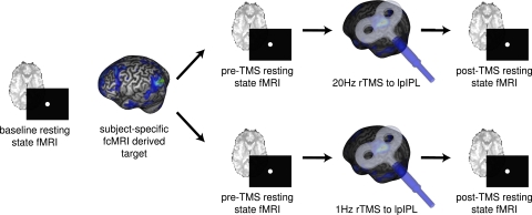
Fig. 1.
Experimental design. Each participant first underwent a baseline resting-state fcMRI scanning session to allow for the creation of individualized functional connectivity maps localizing the lpIPL node of their default network. This node served as the future target for two subsequent rTMS stimulation sessions (on separate days): one in which they received 1-Hz rTMS to lpIPL, and another in which they received 20-Hz rTMS to lpIPL. Consistent targeting of lpIPL across sessions was achieved by using a frameless stereotactic neuronavigation system. During each of the two stimulation sessions, subjects completed two fcMRI scans: one immediately before, and another immediately following rTMS.

