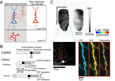
Fig. 1.
Imaging of CaMKII activity in identified OD domains. (A) Schematic diagram of a sample pyramidal neuron (blue) in layer II/III within the deprived eye domain receiving inputs from multiple sources (red). Thick red triangles represent feedforward synapses. Other synapses derive from local and longer-range axons, including inputs from the nondeprived eye. (B) Top: Schematic drawing of the CaMKII protein. Bottom: Conformations of Camui in the inactive and active form. (C) Alignment of two-photon microscopic images with OD map. Blood vessel and OD maps were obtained using intrinsic signal optical imaging (Upper: A, anterior; M, medial). Gray scale indicates ODI (white, ipsilateral eye dominated; black, contralateral eye dominated). Blood vessels in low-magnification optical and two-photon microscopic images were used to align two-photon images (Lower) to OD domains. A dendritic segment (red box) is magnified (Right) and displayed as channel separated images (CFP and YFP) as well as a ratiometric image in intensity-modulated display mode, indicating the CFP/YFP ratio. Warm hue represents high CaMKII activity.

