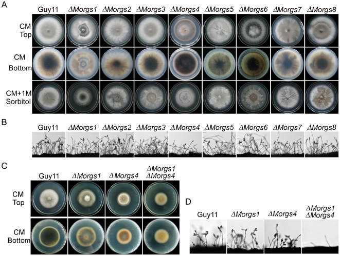
Figure 2. Comparison of various ΔMorgs mutant strains in colony morphology and conidia formation.
(A) Colony morphology was observed by incubating culture plates in the dark for ten days at 28°C and then photographed. (B) Conidia formation was observed under a light microscope 24 hours at room temperature after induction of conidiation under cover slips. (C) Comparison of specific single and double mutants in colony formation in the dark for eight days at 28°C and then photographed. (D) Comparison of specific single and double mutants in conidia formation 24 hours at room temperature after induction of conidiation under cover slips.

