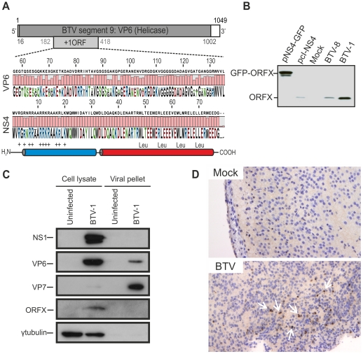
Figure 1. BTV expresses a fourth non structural protein (NS4).
(A) BTV segment 9 (1049 base pairs). The VP6 protein (dark gray) is encoded by nucleotides 16 to 1002. The NS4 coding sequence is located in the +1 open reading frame (ORF) between nucleotides 182 to 418. VP6 (residues 57 to 135) and NS4 (residues 1 to 79) amino acid conservation plots are shown. NS4 secondary structure prediction indicated the presence of two putative α-helices, drawn in blue and red. The N-terminal domain (blue) is highly basic and the C-terminal domain (red) contains a conserved leucine zipper motif. (B) Western blotting of cellular extracts (lysate) of BSR cells either transfected with 1.8 µg of plasmid expressing NS4 alone (pcI-NS4) or in fusion with eGFP (peGFP-NS4), or infected by BTV-8 or BTV-1 at a MOI of 0.01. Cells were analyzed 36 h post-transfection or infection and blots were incubated with NS4 antiserum. (C) Western blots of viral pellets and cell protein extracts of BFAE cells infected by BTV-1 at a MOI of 0.05. Samples were analyzed at 48 h post-infection by SDS-PAGE and western blotting employing antisera against NS1, VP6, VP7, ORFX (NS4) and γ-tubulin as indicated. (D) Immunohistochemical detection of NS4. Immunohistochemistry was performed as described in Materials and Methods in brain tissue sections of mice inoculated with BTV-8 72 h post-infection using an antiserum against NS4. Cells expressing NS4 are stained brown as indicated by white arrows. No expression of NS4 is detected in negative control mice mock-inoculated with cell culture media.

