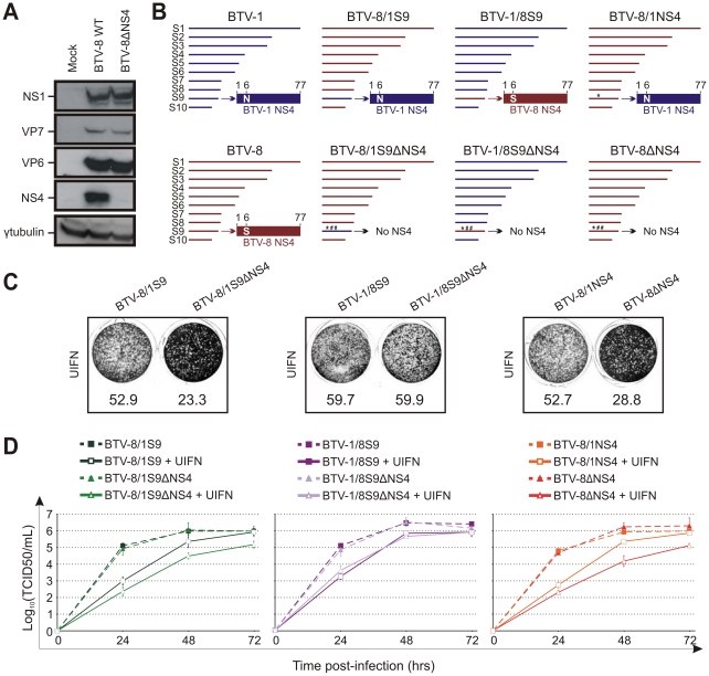
Figure 8. The NS4 of BTV-1 displays similar biological properties to the homologous BTV-8 protein.
(A) Western blotting of cellular extracts (lysate) of CPT-Tert cells infected with BTV-8 wt or BTV-8ΔNS4 at a MOI of 0.01. Cells were analyzed 24 h post-infection and blots were incubated with antisera against NS1, VP7, VP6, NS4 and γ-tubulin as indicated. (B) Schematic diagram of the BTV-8/BTV-1 reassortants and mutants used in this study. Note that BTV1 and BTV8 segments/proteins are coloured in blue and red, respectively. * indicates a point mutation, while # indicates the introduction of a stop codon in the NS4 ORF. (C) CPT-Tert cells were treated with 1000 AVU/ml of Universal IFN for 20 h prior, and 2 h after, being infected by the recombinant viruses indicated in the panel using a MOI of 0.01. Cell monolayers were stained 72 h post-infection using crystal violet. Values indicated below each well correspond to the relative quantification of the disrupted monolayer using Image-Pro Plus (MediaCybernetics, Inc.). (D) CPT-Tert cells were treated (solid line) or mock treated (dashed line) with 100 AVU/ml of Universal interferon (UIFN) for 20 h prior and 2 h after being infected by the viruses indicated in the panel. Cells were infected at a MOI of 0.01. Supernatants were collected at 24, 48 and 72 h after infection, and then titrated on BSR cells by limiting dilution analysis and virus titers expressed as log10 (TCID50/ml). In parallel, each virus preparation was also re-titrated by limiting dilution analysis to control that equal amounts of input virus was used in each experiment. This experiment was performed two times, each time in duplicate.

