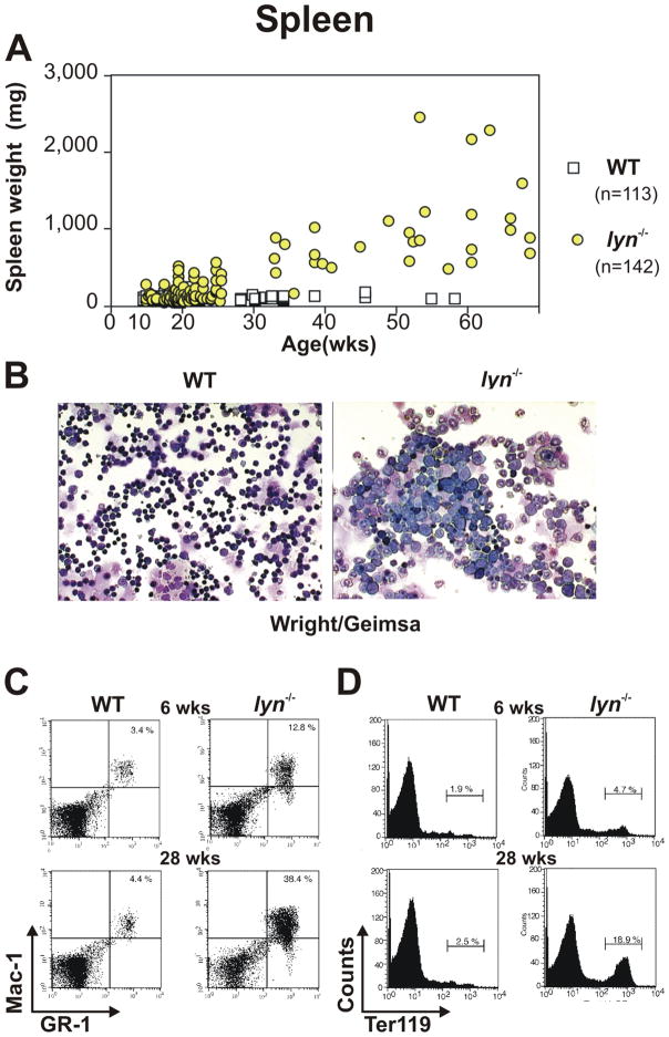
Figure 2. Lyn-deficient mice develop splenomegaly due to myeloproliferation and extra-medullary erythropoiesis.
(A) Spleen weight of WT and lyn−/− mice monitored over 60 weeks. (B) Splenic cells derived from WT and lyn−/− mice stained with Wright/Giemsa. (C, D) Flow cytometric analysis of WT and lyn−/− splenocytes from 6 and 28 week old mice. Percentages of Mac-1/Gr-1 double positive myeloid cells (C) or Ter-119 positive erythroid cells (D) are reported.

