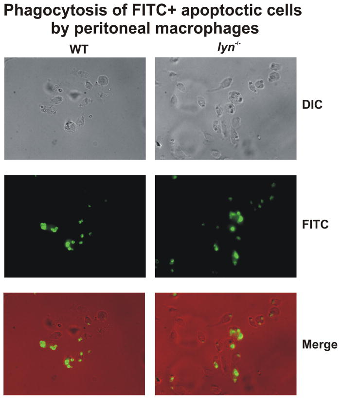
Figure 6. Lyn-deficient macrophages phagocytose apoptotic thymocytes normally.
WT and lyn−/− peritoneal macrophages were collected 5 days after IP injection with aged 3% thioglycollate and cultured overnight. Macrophages were exposed to 20-fold excess of apoptotic thymocytes, labeled with CellTracker green, for 30 min an examined by fluorescence microscopy, as described (108).

