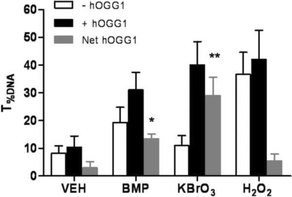
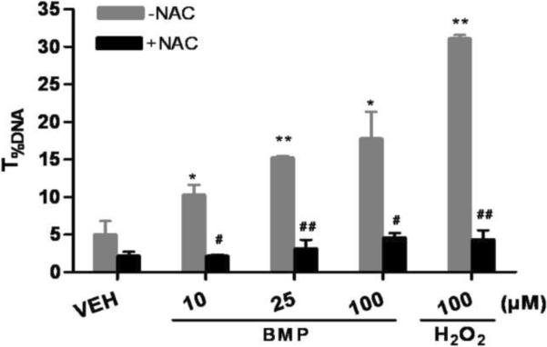
Figure 5.
Involvement of oxidative stress in BMP mediated DNA damage. (A) Oxidative base modifications in UROtsa cells following 1h of BMP (25μM), KBrO3(2mM) and H2O2(100μM) exposure measured by hOGG1 modified comet assay. Gray Bars represent the net increase in T%DNA after incubation with hOGG1. (B) Protective effects of NAC (2mM) on BMP (10–100μM) induced DNA strand breaks in UROtsa cells. Data are shown as mean ± SEM, N=3. *P<0.05 versus vehicle control, #P<0.05 versus cells without NAC pre-treatment.


