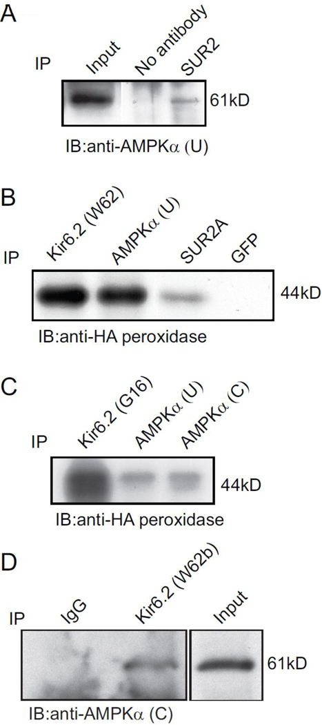
Fig 6.
Co-immunoprecipitation of AMPK α-subunits and KATP channel subunits. Co-immunoprecipitation of cell lysates from cells transfected with Kir6.2-HA and SUR2A cDNAs and rat ventricular tissue. Detection of AMPKα in the immunoprecipitate obtained with anti-SUR2A antibodies. B, Kir6.2-HA protein was detected in the immunoprecipitate obtained with the anti-Kir6.2 (W62), anti-AMPKα (U) and anti-SUR2A antibodies, but not with anti-GFP antibodies that were used as a negative control. C, Detection of Kir6.2-HA protein in immunoprecipitates obtained with a different anti-Kir6.2 (G16) antibody, as well as the immunoprecipitate obtained with two separate anti-AMPKα antibodies. D, Detection of AMPKα subunits in immunoprecipitates obtained with an anti-Kir6.2 (W62b) antibody. An unrelated antibody (rabbit IgG) was used as a negative control. The results shown are representative of 2–3 separate experiments, which showed similar results.

