
Fig. 1.
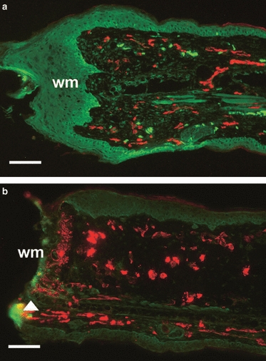
Re-innervation and vascularisation of denervated vs. non-denervated MRL/MpJ ear wounds 7 days post-wounding. Immunohistochemistry assessed the presence of pan-neurofilament (FITC) and CD31 (TRITC) in both non-denervated ear wounds (a) and denervated ear wounds (b) in the MRL/MpJ mouse 7 days post-wounding. (a) Section from the centre of the regenerating ear showing the presence of nerves and blood vessels advancing beyond the cut edge of the cartilage (arrow) and a thickened wound epithelium at the wound margin (wm). (b) Section from the denervated ear showing the denervation procedure was successful as no nerves were present and the blood vessels appear dilated. Note the speckled staining of CD31 (arrow), indicating disintegration of vessels at the necrosing wound margin (wm). Scale bars: 100 μm.