
Fig. 2.
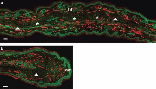
Re-innervation and vascularisation of denervated vs. innervated non-denervated MRL/MpJ ear wounds 50 days post-wounding. Immunohistochemistry was used to detect the presence of neurofilament (FITC) and CD31 (TRITC) in both non-denervated ear wounds (a) and denervated ear wounds (b) in the MRL/MpJ mouse 50 days post-wounding. (a) Section taken from regenerating ear tissue showing cartilage island formation (*), regenerated hair follicles (hf) beyond the original wound margin (wm; arrows). (b) Section from proximal wound margin of a denervated ear showing re-vascularisation, but only a small influx of regenerating nerves between the cut edge of cartilage (arrow) and the wound margin (wm). Scale bars: 100 μm.