
Fig. 5.
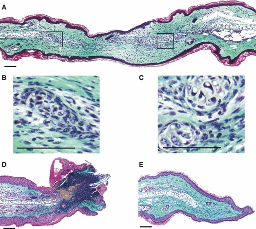
Histology of the denervated and non-denervated MRL/MpJ ear wounds 50 days post-wounding. Masson's Trichrome stained tissue sections. Non-denervated ear: (A) section through the centre of the regenerating wound displaying collagen deposition, epithelial fusion of opposing wound margins, and chondrogenesis with the formation of numerous cartilage islands (magnified in B and C). Denervated ear: (D) section through the distal edge of the wound at the necrosing ear tip, displaying severe regression of the wound with necrosis and failure to re-epithelialise; (E) section through the proximal wound edge showing re-epithelialisation, collagen deposition and proliferation at the cartilage stump (arrow). However, no characteristic features of a regenerative blastema-like structure were observed in the denervated MRL/MpJ ear wound. Scale bars: 100 μm.